20583741
Descripción
Test por Charlotte Jakes, actualizado hace más de 1 año
|
|
Creado por Charlotte Jakes
hace más de 4 años
|
|
Pregunta 1
Pregunta
What range of flexion can the C spine achieve?
Respuesta
-
40 degrees
-
60 degrees
-
20 degrees
-
35 degrees
Pregunta 2
Pregunta
What range of extension can the C spine achieve?
Respuesta
-
40 degrees
-
60 degrees
-
20 degrees
-
10 degrees
Pregunta 3
Pregunta
What range of flexion can the T spine achieve?
Respuesta
-
35 degrees
-
20 degrees
-
40 degrees
-
60 degrees
Pregunta 4
Pregunta
What range of extension can the T spine achieve?
Respuesta
-
20 degrees
-
35 degrees
-
60 degrees
-
40 degrees
Pregunta 5
Pregunta
What range of extension can the L spine achieve?
Respuesta
-
25 degrees
-
60 degrees
-
20 degrees
-
40 degrees
Pregunta 6
Pregunta
What range of flexion can the L spine achieve?
Respuesta
-
60 degrees
-
25 degrees
-
35 degrees
-
40 degrees
Pregunta 7
Pregunta
What is the total range of flexion that the vertebral column can achieve?
Respuesta
-
250 degrees
-
110 degrees
-
140 degrees
-
100 degrees
Pregunta 8
Pregunta
What range of lateral flexion can the L spine achieve?
Respuesta
-
20 degrees
-
35 degrees
-
60 degrees
Pregunta 9
Pregunta
What range of lateral flexion can the T spine achieve?
Respuesta
-
20 degrees
-
35 degrees
-
60 degrees
Pregunta 10
Pregunta
What range of lateral extension can the C spine achieve?
Respuesta
-
20 degrees
-
35 degrees
-
60 degrees
Pregunta 11
Pregunta
What degree of rotation can be achieved around the sacrum and lumbar spine?
Respuesta
-
5 degrees
-
35 degrees
-
50 degrees
Pregunta 12
Pregunta
What degree of rotation can be achieved around the lumbar and thoracic spine?
Respuesta
-
35 degrees
-
5 degrees
-
50 degrees
Pregunta 13
Pregunta
What degree of rotation can be achieved around the cervical and thoracic spine?
Respuesta
-
50 degrees
-
35 degrees
-
5 degrees
Pregunta 14
Pregunta
What degree of rotation can be achieved by the entire vertebral column?
Respuesta
-
90-95 degrees
-
60-65 degrees
-
100-105 degrees
-
70-75 degrees
Pregunta 15
Pregunta
How do we classify the intervertebral discs?
Respuesta
-
Secondary cartilaginous joints
-
Primary cartilaginous joints
-
Fibrous joint
-
Synovial joint
Pregunta 16
Pregunta
What do the intervertebral discs allow that would not be possible if the flat articular surfaces of the vertebrae were joined directly?
Respuesta
-
Rocking
-
Rotations
-
Abduction
-
Flexion
Pregunta 17
Pregunta
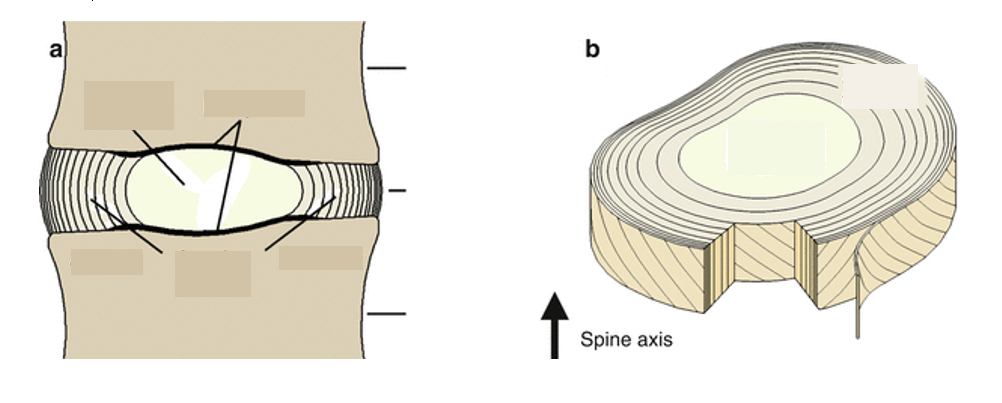
Label these two diagrams of the intervertebral disc.
Respuesta
-
Nucleus pulposus
-
Annulus fibrosus
-
End plate
-
Body
-
Body
-
Nucleus pulposus
-
Annulus fibrosus
Pregunta 18
Pregunta
What type of collagen will you find in the nucleus pulposus and annulus fibrosus?
Respuesta
-
Type I
-
Type II
-
Type IV
-
Type X
Pregunta 19
Pregunta
How is the nucleus pulposus adapted for weight-bearing?
Respuesta
-
High water content resists compression
-
Collagen type I resists compression
-
High amino acid content resists compression
-
Formed of tough fibrous layers
Pregunta 20
Pregunta
Which is true of the annulus fibrosus?
Respuesta
-
Thicker anteriorly and more tightly packed posteriorly
-
More tightly packed anteriorly and thicker posteriorly
-
Evenly packed over the entire vertebral body
-
Even thickness over the entire vertebral body
Pregunta 21
Pregunta
Why is the annulus fibrosus an efficient shock absorber?
Respuesta
-
Each layer has fibres in a different orientation to the last - changes in angulation cause slow deformation
-
Each layer has fibres in the same orientation - aligned angulation causes slow deformation
-
Consists of collagen type II which is flexible
-
High water content to resist compression
Pregunta 22
Pregunta
What is the vertebral end plate?
Respuesta
-
The point of fusion of the intervertebral disc with the vertebral body
-
The gel-like central portion of the intervertebral disc
-
The layers of tough fibrous tissue forming the bulk of the intervertebral disc
-
The point of fusion of the intervertebral disc with the spinous process
Pregunta 23
Pregunta
What happens in a herniated disc?
Respuesta
-
The nucleus pulposus protrudes into the annulus fibrosus
-
The annulus fibroses extends behind the vertebral body
-
The nucleus pulposus protrudes into the vertebral foramen
-
The vertebral end plate breaks down
Pregunta 24
Pregunta
What is the vertebral end plate formed of?
Respuesta
-
Hyaline cartilage
-
Collagen type I
-
Elastic tissue
-
Smooth muscle
Pregunta 25
Pregunta
Through which component of the intervertebral discs are water and nutrients received?
Respuesta
-
Vertebral end plate
-
Annulus fibrosus
-
Nucleus pulposus
Pregunta 26
Pregunta
Why do the intervertebral discs become thinner and the flexibility of the vertebral column decrease with age?
Respuesta
-
Decline in water content of nucleus pulposus
-
Loss of angulation of fibres of annulus fibrosus
-
Increase in water content of nucleus pulposus
-
Degradation of the vertebral end plate
Pregunta 27
Pregunta
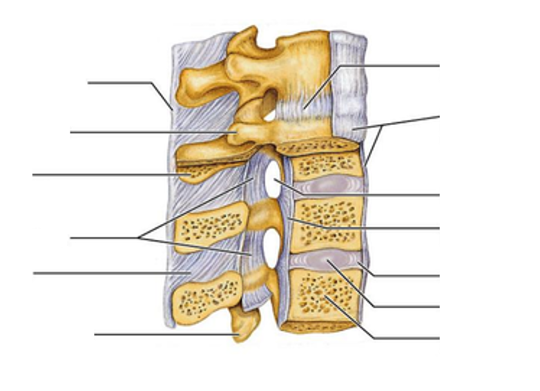
Label this image to show the ligaments of the vertebral column.
Respuesta
-
Posterior longitudinal ligament
-
Anterior longitudinal ligament
-
Ligamentum flavum
-
Ligamentum flavum
-
Supraspinous ligament
-
Interspinous ligament
-
Supraspinous ligament
Pregunta 28
Pregunta
The posterior longitudinal ligament lines the inside of the vertebral canal.
Respuesta
- True
- False
Pregunta 29
Pregunta
Where does the ligamentum flavum occur?
Respuesta
-
Between the lamina of adjacent vertebrae
-
Between the pedicles of adjacent vertebrae
-
Along the apex of the spinous processes
-
Along the anterior side of the vertebral bodies
Pregunta 30
Pregunta
The nuchal ligament is an extension of what up the cervical spine?
Respuesta
-
Supraspinous ligament
-
Posterior longitudinal ligament
-
Anterior longitudinal ligament
-
Intervertebral discs
Pregunta 31
Pregunta
Label this image of the vertebral column and its ligaments.
Respuesta
-
Supraspinous ligament
-
Transverse process
-
Spinous process
-
Ligamentum flavum
-
Interspinous ligament
-
Inferior articular process
-
Body
-
Nucleus pulposus
-
Annulus fibrosus
-
Posterior longitudinal ligament
-
Intervertebral foramina
-
Anterior longitudinal ligament
-
Intervertebral disc
Pregunta 32
Pregunta
Fill in the blanks below to describe the muscles of the trunk.
The muscles of the posterior abdominal wall are [blank_start]extensors[blank_end].
The muscles that line the lateral and anterior abdominal wall are responsible for f[blank_start]lexion[blank_end] and r[blank_start]otation[blank_end].
The [blank_start]superficial[blank_end] posterior trunk muscles are mostly responsible for the movement of the shoulder.
The [blank_start]intermediate[blank_end] posterior trunk muscles are mostly responsible for the movement and stability of the thoracic cage.
The [blank_start]deep[blank_end] posterior trunk muscles are mostly responsible for the stabilisation of the vertebral column.
Respuesta
-
extensors
-
lexion
-
otation
-
superficial
-
intermediate
-
deep
Pregunta 33
Pregunta
The erector-spinae muscles form part of the [blank_start]deep[blank_end] back muscles. They consist of the i[blank_start]liocoastalis[blank_end], l[blank_start]ogissimus[blank_end] and s[blank_start]pinalis[blank_end] muscles. They extend and laterally [blank_start]rotate[blank_end] the trunk as well as maintain correct [blank_start]posture[blank_end].
Respuesta
-
deep
-
liocoastalis
-
ogissimus
-
pinalis
-
rotate
-
posture
Pregunta 34
Pregunta
The transversospinalis muscles form a part of the [blank_start]deep[blank_end] muscles of the back. They consist of the r[blank_start]otatores[blank_end], the m[blank_start]ultifidus[blank_end] and the s[blank_start]emispinalis[blank_end] muscles. They lie between the t[blank_start]ransverse[blank_end] and s[blank_start]pinous[blank_end] processes of the vertebrae to rotate and [blank_start]extend[blank_end] the vertebral column.
Respuesta
-
deep
-
otatores
-
ultifidus
-
emispinalis
-
ransverse
-
pinous
-
extend
Pregunta 35
Pregunta
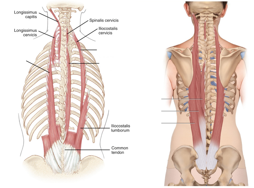
Fill in the blanks to label the erector spinae group of deep back muscles.
Respuesta
-
Longissimus
-
Spinalis
-
Iliocostalis
-
Iliocostalis
-
Longissimus
-
Spinalis
Pregunta 36
Pregunta
Fill in the blanks below to label the transversospinalis group of muscles.
Respuesta
-
Multifidus
-
Interspinalis
-
Semispinalis
-
Rotatores
-
Multifidus
-
Rotatores
Pregunta 37
Pregunta
Fill in the blanks below to describe the intermediate back muscles.
The serratus posterior superior muscle originates from the [blank_start]nuchal[blank_end] ligament as well as the spinous processes of C[blank_start]7[blank_end]-T[blank_start]3[blank_end]. It inserts at the superior borders of the [blank_start]2[blank_end]nd-[blank_start]5[blank_end]th ribs.
The serratus posterior inferior muscle originates from the spinous processes of T[blank_start]11[blank_end]-L[blank_start]2[blank_end] and inserts at the inferior borders of the [blank_start]9[blank_end]th-[blank_start]12[blank_end]th ribs.
Respuesta
-
nuchal
-
7
-
3
-
2
-
5
-
11
-
2
-
9
-
12
Pregunta 38
Pregunta
Which is true of the serratus posterior muscles?
Respuesta
-
Both elevate the ribs
-
Both depress the ribs
-
The serratus posterior superior muscle elevates the ribs whilst the serratus posterior inferior muscle depresses the ribs
-
The serratus posterior superior muscle depresses the ribs whilst the serratus posterior inferior muscle elevates the ribs
Pregunta 39
Pregunta
Label the image below to show the intermediate muscles of the back.
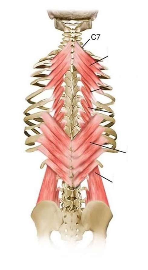
Respuesta
-
Serratus posterior superior
-
Serratus posterior inferior
-
Quadratus lumborum
Pregunta 40
Pregunta
Fill in the blanks to label this image of the superficial back muscles.
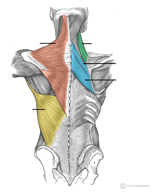
Respuesta
-
Trapezius
-
Latissimus dorsi
-
Rhomboid major
-
Rhomboid minor
-
Levator scapulae
Pregunta 41
Pregunta
Fill in the blanks to describe the origins and insertions of the superficial muscles of the back.
The trapezius originates from the s[blank_start]kull[blank_end], ligamentum [blank_start]nuchae[blank_end] and spinous processes of C[blank_start]7[blank_end]-T[blank_start]12[blank_end] to insert at the c[blank_start]lavicle[blank_end], a[blank_start]cromion[blank_end] and s[blank_start]capula[blank_end] spine.
The latissimus dorsi originates from the [blank_start]thoracolumbar[blank_end] fascia at T[blank_start]6[blank_end]-T[blank_start]12[blank_end], the [blank_start]iliac[blank_end] crest and the most inferior [blank_start]3[blank_end] ribs to insert at the intertubular sulcus of the [blank_start]humerus[blank_end].
The levator scapulae originates from the transverse processes of C[blank_start]1[blank_end]-C[blank_start]4[blank_end] and inserts at the s[blank_start]capula[blank_end].
Rhomboid minor originates from the spinous processes of C[blank_start]7[blank_end]-T[blank_start]1[blank_end] and inserts at the s[blank_start]capula[blank_end].
Rhomboid major originates from the spinous processes of T[blank_start]2[blank_end]-T[blank_start]5[blank_end] and inserts at the s[blank_start]capula[blank_end].
Respuesta
-
kull
-
nuchae
-
7
-
12
-
lavicle
-
cromion
-
capula
-
thoracolumbar
-
6
-
12
-
iliac
-
3
-
humerus
-
1
-
4
-
capula
-
7
-
1
-
capula
-
2
-
5
-
capula
Pregunta 42
Pregunta
What innervates the trapezius?
Respuesta
-
Accessory cranial nerve XI
-
Thoracodorsal nerve
-
Dorsal scapula nerve
Pregunta 43
Pregunta
What innervates the latissimus dorsi?
Respuesta
-
Accessory cranial nerve XI
-
Thoracodorsal nerve
-
Dorsal scapula nerve
Pregunta 44
Pregunta
Which nerve innervates levator scapulae?
Respuesta
-
Dorsal scapula nerve
-
Accessory cranial nerve XI
-
Thoracodorsal nerve
Pregunta 45
Pregunta
Which nerve innervates both rhomboid minor and rhomboid major?
Respuesta
-
Dorsal scapula nerve
-
Accessory cranial nerve XI
-
Thoracodorsal nerve
Pregunta 46
Pregunta
Which superifical back muscle elevates, retracts and depresses the scapula?
Respuesta
-
Trapezius
-
Latissimus dorsi
-
Levator scapula
-
Rhomboids
Pregunta 47
Pregunta
Which superficial back muscle aducts, extends and medially rotates the upper arm?
Respuesta
-
Latissimus dorsi
-
Trapezius
-
Levator scapulae
-
Rhomboids
Pregunta 48
Pregunta
Which superficial back muscle pnly elevates the scapula?
Respuesta
-
Trapezius
-
Latissimus dorsi
-
Rhomboids
-
Levator scapulae
Pregunta 49
Pregunta
Which superficial back muscle rotates and retracts the scapula?
Respuesta
-
Rhomboids
-
Levator scapulae
-
Latissimus dorsi
-
Trapezius
Pregunta 50
Pregunta
Where is the thoracolumbar fascia inserted?
Respuesta
-
The hip bones and sacrum
-
The sacrum and coccyx
-
The iliac crests
-
The pubic tubercles
Pregunta 51
Pregunta
Label this image to show the relations of the thoracolumbar fascia in the back.
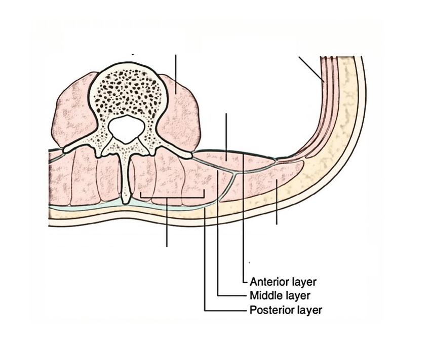
Respuesta
-
Thoracolumbar fascia
-
Erector spinae
-
Psoas major
-
Quadratus lumborum
-
Latissimus dorsi
-
Transversus abdominis
Pregunta 52
Pregunta
When does the thoracolumbar fascia tense?
Respuesta
-
When the muscles of the back contract
-
When the anterior abdominal wall muscles contract
-
When the muscles of the back relax
-
When the vertebral column is in flexion
Pregunta 53
Pregunta
Label this diagram to show the muscles of the back.
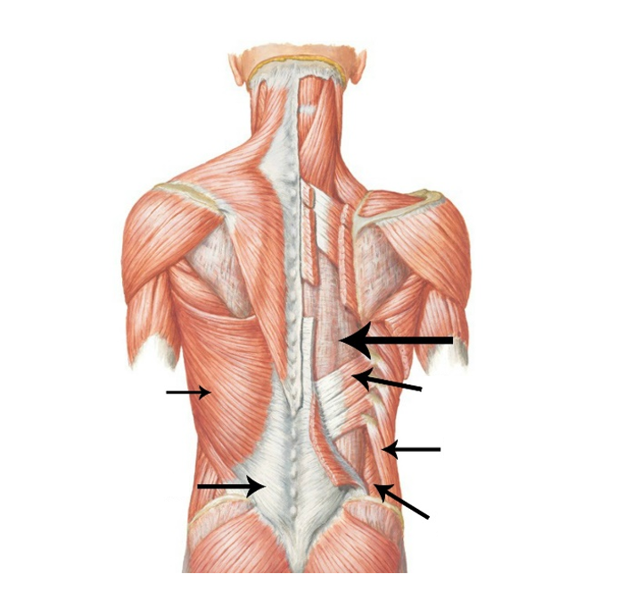
Respuesta
-
Erector spinae
-
Latissimus dorsi
-
Thoracolumbar fascia
-
Internal oblique
-
External oblique
-
Serratus posterior inferior
Pregunta 54
Pregunta
Fill in the blanks below to describe the origin and insertions of the lateral trunk muscles.
Quadratus lumborum originates from the i[blank_start]liac crest[blank_end]. It inserts onto the [blank_start]transverse[blank_end] processes of L[blank_start]1[blank_end]-L[blank_start]4[blank_end] and the inferior border of the [blank_start]12[blank_end]th rib.
Iliacus originates from the surface of the iliac [blank_start]fossa[blank_end] and [blank_start]anterior inferior[blank_end] iliac spine. It inserts at the [blank_start]lesser trochanter[blank_end] of the [blank_start]femur[blank_end] in combination with [blank_start]psoas major[blank_end].
Psoas minor, when present, originates from the vertebral [blank_start]bodies[blank_end] of T[blank_start]12[blank_end] and L[blank_start]1[blank_end] and inserts at the [blank_start]pectineal[blank_end] line of the [blank_start]pubis[blank_end].
Psoas major originates from the [blank_start]transverse[blank_end] processes and vertebral bodies of T[blank_start]12[blank_end]-L[blank_start]5[blank_end] and inserts at the [blank_start]lesser trochanter[blank_end] of the femur.
Respuesta
-
liac crest
-
1
-
transverse
-
4
-
12
-
fossa
-
anterior inferior
-
lesser trochanter
-
femur
-
psoas major
-
12
-
1
-
pectineal
-
pubis
-
12
-
5
-
transverse
-
bodies
-
lesser trochanter
Pregunta 55
Pregunta
Psoas minor inserts at the lesser trochanter of the femur.
Respuesta
- True
- False
¿Quieres crear tus propios Tests gratis con GoConqr? Más información.