20655810
Descripción
Test por Charlotte Jakes, actualizado hace más de 1 año
|
|
Creado por Charlotte Jakes
hace más de 4 años
|
|
Pregunta 1
Pregunta
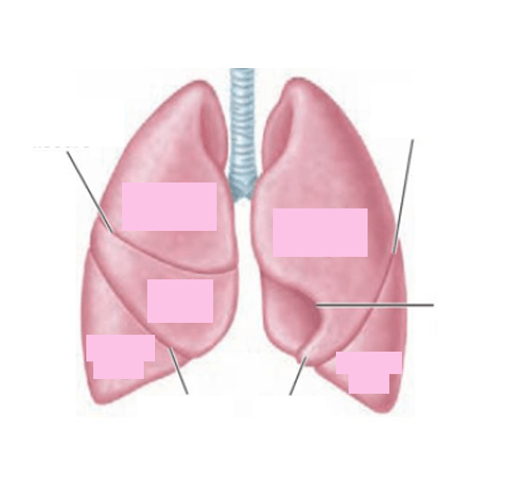
Label the diagram to show the lobes and fissures of the lungs.
Respuesta
-
Horizonal fissure
-
Superior lobe
-
Middle lobe
-
Inferior lobe
-
Superior lobe
-
Inferior lobe
-
Oblique fissure
-
Oblique fissure
-
Lingula
-
Cardiac notch
Pregunta 2
Pregunta
The right lung is slightly larger than the left.
Respuesta
- True
- False
Pregunta 3
Pregunta
Where is the lingula in relation to the heart?
Respuesta
-
Loops around the left border
-
Loops around the aortic arch
-
Deep to the inferior vena cava
-
Loops around the apex
Pregunta 4
Pregunta
How many lobes does the right lung have?
Respuesta
-
3
-
2
-
1
-
4
Pregunta 5
Pregunta
How many lobes does the left lung have?
Respuesta
-
1
-
2
-
3
-
4
Pregunta 6
Pregunta
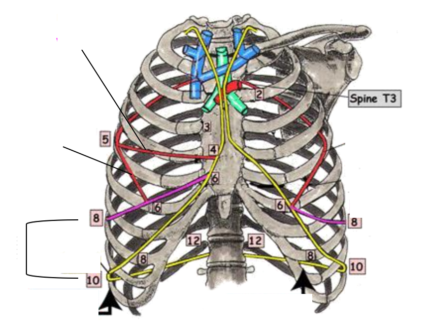
Drag and drop the correct answers to describe the orientation of the lubes of the lungs.
The left and right oblique fissures run from the spinous process of [blank_start]T3[blank_end] to the [blank_start]6th rib[blank_end] at the [blank_start]midclavicular line[blank_end].
The right horizontal fissure runs from the [blank_start]5th rib[blank_end] at the [blank_start]midaxillary line[blank_end] and the [blank_start]4th rib[blank_end].
Respuesta
-
T3
-
T1
-
T2
-
6th rib
-
7th rib
-
3rd rib
-
midclavicular line
-
5th rib
-
midaxillary line
-
4th rib
Pregunta 7
Pregunta
Why can PET scans show lung masses?
Respuesta
-
Show metabolic activity
-
Lung masses will be formed of dense tissue that absorbs X ray
-
Lung masses will be formed of dense tissue that absorbs gamma
-
Lung masses will be formed of dense tissue that has resonance
Pregunta 8
Pregunta
What is a pneumothorax?
Respuesta
-
Puncture of the pleural cavity causing abnormal movements of air
-
Excess fluid in the pleural cavity due to infection
-
Excess fluid in the pericardial cavity
-
Blood in the pericardial cavity
Pregunta 9
Pregunta
What will happen to air in a pneumothorax during expiration?
Respuesta
-
Air leaves pleural cavity
-
Air enters pleural cavity
Pregunta 10
Pregunta
What will happen to air in a pneumothorax during inspiration?
Respuesta
-
Leaves the pleural cavity
-
Enters the pleural cavity
Pregunta 11
Pregunta
Fill in the blanks to describe a tension pneumothorax.
A tension pneumothorax occurs when the defect in the chest wall acts as a flap [blank_start]valve[blank_end]. During [blank_start]expiration[blank_end], the defect [blank_start]closes[blank_end] and air cannot leave the thorax. During [blank_start]inspiration[blank_end], great pressure caused by the pleural cavity filling with [blank_start]air[blank_end] causes the heart to be shifted to the right. This compresses the heart and lung.
Respuesta
-
valve
-
closes
-
expiration
-
inspiration
-
air
Pregunta 12
Pregunta
What name do we have the harsh transition between misty grey tissue and black tissue on an X-ray of a pneumothorax?
Respuesta
-
Pleural stripe
-
Pleural margin
-
Pleural edge
-
Pleural boundary
Pregunta 13
Pregunta
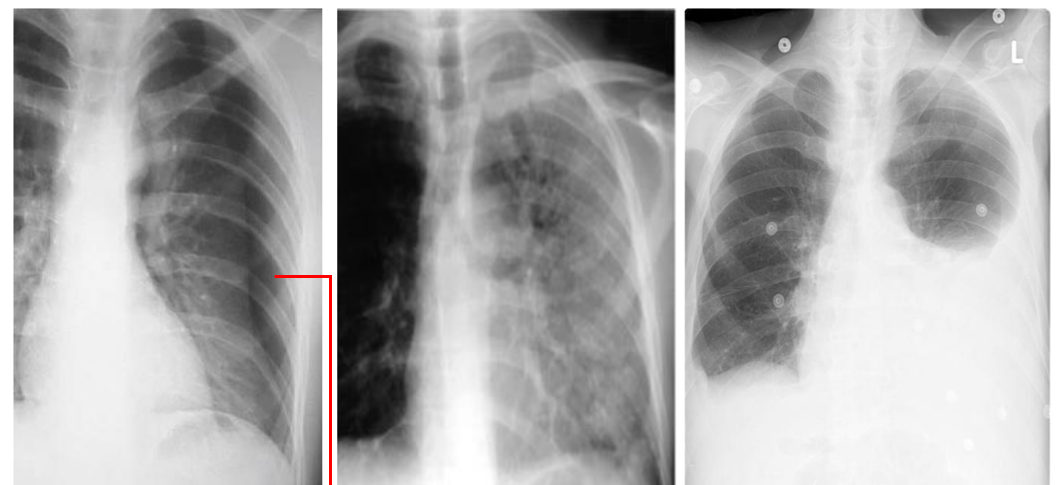
Drag and drop the correct lung pathology to its x ray.
Respuesta
-
Pneumothorax
-
Pneumonia
-
Pleural effusion
Pregunta 14
Pregunta
Why is there consolidation of lung tissue in x-rays of chests with pneumonia?
Respuesta
-
Accumulation of lymphocytes causes fluid buildup
-
Irritants in pleura stimulate serous membranes to produce extra fluid
-
Air in the pleural cavity appears misty grey
-
Accumulation of pus
Pregunta 15
Pregunta
What is pleural effusion?
Respuesta
-
Irritants in the parietal pleura stimulate serous membranes to produce extra fluid
-
Accumulation of lymphocytes causes fluid buildup
-
Irritants in parietal pleura cause serous membranes to stop producing serous fluid
-
Air in the pleural cavity due to puncture of the parietal pleura
Pregunta 16
Pregunta
What is the terminal functional unit of the lung?
Respuesta
-
Bronchopulmonary segment
-
Lobe
-
Bronchiole
-
Alveolus
Pregunta 17
Pregunta
What is the correct orientation of the hilum of the right lung, superior to inferior?
Respuesta
-
Bronchus, artery, vein
-
Artery, bronchus, vein
-
Vein, artery, bronchus
-
Vein, bronchus, artery
Pregunta 18
Pregunta
What is the correct orientation of the lung hilum of the left lung, superior to inferior?
Respuesta
-
Artery, bronchus, vein
-
Bronchus, vein, artery
-
Bronchus, artery, vein
-
Vein, bronchus, artery
Pregunta 19
Pregunta
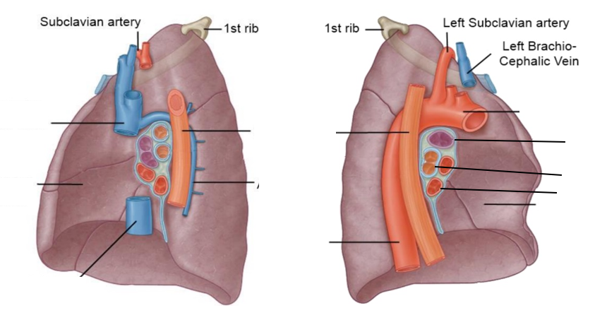
Label this image of the posterior surfaces of the lungs and their relations.
Respuesta
-
Superior vena cava
-
Cardiac notch
-
Inferior vena cava
-
Azygos vein
-
Oesophagus
-
Oesophagus
-
Descending aorta
-
Aortic arch
-
Pulmonary artery
-
Bronchus
-
Pulmonary vein
-
Cardiac notch
Pregunta 20
Pregunta
The bronchus starts dividing in the hilum of the lung at the root.
Respuesta
- True
- False
Pregunta 21
Pregunta
Label this image to show the positions of the structures of the lungs.
Image:
Lungs (binary/octet-stream)
Respuesta
-
Left oblique fissure
-
Visceral pleura
-
Parietal pleura
-
Costodiaphragmatic recess
-
Right oblique fissure
-
Right horizontal fissure
-
Midaxillary line
-
Midclavicular line
Pregunta 22
Pregunta
Drag the correct answers to describe how you would perform auscultation on the anterior chest wall.
To listen to the apex of the lung you would listen [blank_start]above the clavicle[blank_end].
To listen to the superior lobe of the right lung you would listen [blank_start]in the 2nd intercostal space[blank_end].
To listen to the middle lobe of the right lung you would listen [blank_start]in the 4th intercostal space[blank_end].
To listen to the inferior lobe of the right lung you would listen [blank_start]in the 6th intercostal space[blank_end].
Respuesta
-
above the clavicle
-
in the 2nd intercostal space
-
in the 3rd intercostal space
-
in the 4th intercostal space
-
in the 5th intercostal space
-
in the 6th intercostal space
-
in the 7th intercostal space
Pregunta 23
Pregunta
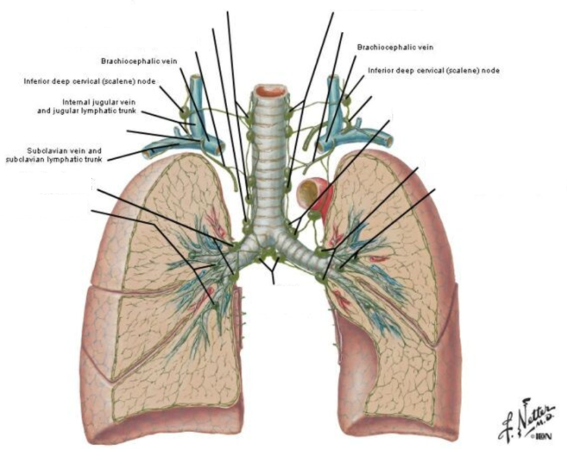
Label this image to show the lymph drainage of the lungs.
Respuesta
-
Bronchopulmonary nodes
-
Intrapulmonary nodes
-
Intrapulmonary nodes
-
Inferior tracheobronchial nodes
-
Bronchopulmonary nodes
-
Left superior tracheobronchial nodes
-
Right superior tracheobronchial nodes
-
Bronchomediastinal lymphatic trunk
-
Right paratracheal nodes
-
Left paratracheal nodes
-
Bronchomediastinal lymphatic trunk
-
Right lymphatic duct
-
Thoracic duct
Pregunta 24
Pregunta
Which of the pleural membranes are supplied by branches of segmental intercostal nerves from the thoracic vertebrae?
Respuesta
-
Cervical
-
Costal
-
Diaphragmatic
-
Mediastinal
Pregunta 25
Pregunta
Which of the pleural membranes are innervated by the phrenic nerves?
Respuesta
-
Costal
-
Cervical
-
Diaphragmatic
-
Mediastinal
Pregunta 26
Pregunta
Where is pain more highly localised?
Respuesta
-
Costal and cervical pleura
-
Diaphragmatic and mediastinal pleura
Pregunta 27
Pregunta
Where is pain referred to the shoulders?
Respuesta
-
Diaphragmatic and mediastinal
-
Costal and cervical
Pregunta 28
Pregunta
What is the parasympathetic innervation of the lungs from?
Respuesta
-
Vagus
-
Accessory cranial nerve
-
T1-T4/5
-
Phrenic nerves
Pregunta 29
Pregunta
What is the sympathetic innervation of the lungs from?
Respuesta
-
T1-T4/5
-
Phrenic nerves
-
Vagus nerve
-
Accessory cranial nerve
Pregunta 30
Pregunta
Parasympathetic stimulation causes bronchoconstriction.
Respuesta
- True
- False
Pregunta 31
Pregunta
Sympathetic stimulation causes bronchodilatation.
Respuesta
- True
- False
Pregunta 32
Pregunta
The sympathetic and parasympathetic nerves that innervate the lungs form anterior and posterior plexuses.
Respuesta
- True
- False
¿Quieres crear tus propios Tests gratis con GoConqr? Más información.